Intervertebral discs are they jelly donuts
- Home
- Blog
Human Intervertebal Disc
A blog on the human intervertebral disc, movement, function, adaptation, degradation,
degeneration, wedding
photos, boiling frogs and Jelly Donut lies.
This blog is on all things about the human intervertebral disc and it’s beautiful structure
which allows it to
form multiple parts of the vertebral column primarily allowing motion of the protective
column and transition of
forces from the lower and upper limbs.
Firstly let’s get some myths out of the way. Human discs do not slip, they really aren’t
“discs”, they are
nothing like a Jelly (jam) donut or a rubber washer, they do not exist in isolation and are
made up of connective
tissues of a predominantly cartilaginous nature. Thus their form is determined by their
function and the ability
of their cellular components to secrete extracellular material. They are living adaptable
structures blending
from the bone and endplate cartilage, they are intimately connected to both, forming a solid
matrix joint
allowing motion and load/force transfer. They work functionally in conjunction with their
zygapophyseal joints to
produce motion and stability to the vertebral column.
It is important to remember the lumbar discs are structurally different than the thoracic
and cervical discs,
they represent the classic annulus and nucleus seen in most text books. The cervical discs
start out similar
looking to lumbar discs but by the time we are adults they have remodelled into a more
crescent shaped
cartilaginous structure without a distinguishable annular nuclear arrangement as in the
lumbar spine
(1)
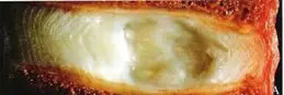
Fig: Section through a disc
Moving forward I will just focus on the lumbar intervertebral joint, and to be more precise
the “interbody
joint” which has colloquially become known as “the disc”.
The interbody joint is different from synovial joints and as a symphesis joint it has a
solid construct with
grading of it’s connective tissue structure based on the local joint internal forces and
function. The outer
capsule of the joint is the outer annulus which is fibrocartilagenous in nature,has a good
vascular and nerve
supply, like all joint capsules. The annulus is made up of fiberous collagen which has
alternating angular fibers
which producing a robust ligamentous structure that is ideal for resisting tensile forces.
As you approach the
inner joint the annulus blends into the nucleus which is high in Glycosaminoglycans (GAGs),
hydrophilic (water
attracting) in nature which give the nucleus (a slolid) the ability to resist compression
and allows axial joint
compression forces to be distributed to the annulus and endplate (2)
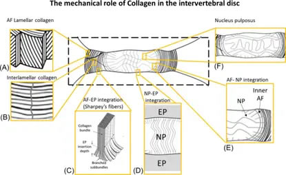
The Mechanical Role of Collagen Fibers in the Intervertebral Disc
The interbody joint (IV disc) is a living joint whose cells require nutrition to maintain
homeostasis and the
joints structure. The inner part of the joint (nucleus) is avascular and gets it’s nutrition
from diffusion
through the end plate and a small amount via the circulation of the outer annulus (3)
Thus movement, loading and then unloading of the joints are required to allow diffusion of
oxygen and nutrients.
The interbody joint, like all cartilage joint structures, requires movement and loading for
life and health.
So to summarise the interbody joints are perfect for their function of load transfer and
movement, they are one
with the bones and endplate not separate enteties. They have a nerve supply and blood supply
in the outer third
and get their internal nutrition from diffusion. They are not rubber washers or anything
like jelly donuts, they
are a perfectly adapted hydraulic joint.
The interbody joints, due to their innervation, are a possible source of pain (nocicpetion)
like all human
joints. Much is written about the pathology that affect the joint from bulging, herniations
to the dreaded
“degenerative disc disease”, touted as a possible mechanism for chronic back pain. The
problem is that
degeneration of the interbody joint is common both in asymptomatic and symptomatic
individuals (although there is
a higher prevelance and correlation in people with symptoms in symptomatic people (4)
Degenerative changes tend to follow a regular course starting with degradation in the
nucleus and progressing to
the annulus (5)

The conundrum is that these changes are common in all age groups even adolescents (6) and
their prevalence
increase with age, but again are common in asymptomatic individuals (7)
So why do some people experience pain and others don’t?
I propose the “Boiling Frog” hypothesis (see below)
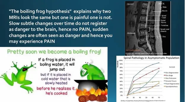
I would also propose that the evidence for degenerative changes are that inherited genetics,
occupational load
exposure, sport exposure and lifestyle have moderate effect on the prevalence (8)
It is actually normal to have degenerative changes in the interbody joint as you age, it is
also normal to have
no symptoms. This would suggest that based on the Boiling Frog Hypothesis above , the
changes seen on MRI scans
in differing age groups are Normal Age (Activity) Related Adaptive Changes (NARAC) not a
pathology or
disease.
The reason that some people develop symptoms is probably also related to the rule of TOOs,
both physical and
psychosocial loads, as shown in the model below.

There inter-body joint can be strained and damaged under sudden or repetitive overload, like all joints in the human body. If the stress and load applied is within the capacity of the joint to remodel and adapt then no symptoms will ensue. There is evidence that the joint will adapt following Wolff’s law like most if not all connective tissue predominant structures (9)

Degenerative changes on MRI may best be viewed as normal changes that develop over time than
a disease or
serious pathology. Due to the high incidence of so called “pathology” on the MRI scans of
asymptomatic
individuals I propose that the Wedding Photo model be used.
In conclusion, it may be normal to have degenerative changes on an MRI. There is some
evidence that people with
pain do have a higher incidence of degenerative findings, but we still do not know why. I
propose that it is the
development and mechanisms that produce the “pathology” on MRI which may lead to some people
to develop symptoms
whilst others don’t. Overloading the joint, which is innervated, may lead to a nociceptive
event causing pain.
The boiling frog model explains this, whilst using the wedding photo analogy will allow
clinicians to rely on
their patients history and examination not their scan results to formulate a diagnosis and
treatment plan.
Beware of VOMIT Victims Of Modern Imaging Technology (10)
Remember degenerative changes are adaptation not pathology per se, MRIs can’t see pain, only
the patients can
report this.